OPM Data processing in Fiji
OPM Data
The data generated by the OPM is in TIFF format. Whether you acquire a single image or a z-stack, you will get a single TIFF file containing all the images.
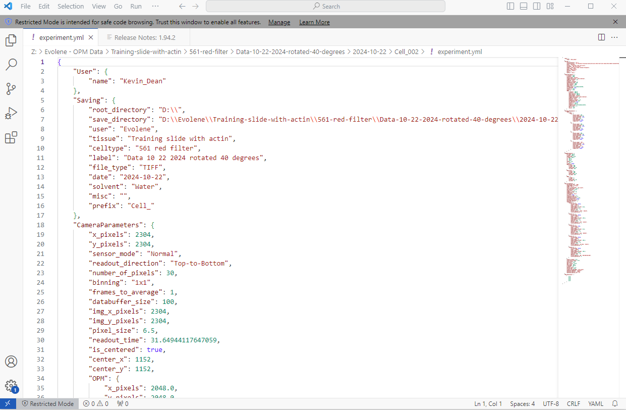
The software also saves an "experiment" file in Yaml format that contains all the experiment settings as well as a "waveform_constants" file in Yaml format that contains the waveform settings. These files can be opened by different applications such as notepad, etc. If you have Visual Studio on your computer, it is a great application to open the file in a structured way. See example below.
Data processing with Fiji/ImageJ
Of course, you are welcome to use any software/method you like to process, analyze and render your data. The TIFF file format should make it easy to open by most softwares.
However, until further development of the instrument, the acquired data is skewed due to the angle of the beam scanning to capture the z-stack. Before visualizing it, you will need to perform a shearing operation to "de-skew" the data.
THE BECKMAN CENTER STAFF WILL DO THIS FOR YOU!!
But if you are curious about how to do it yourself, here are the steps:
There is a way to do this using Fiji/ImageJ. It requires a GPU and CLIJ installed on ImageJ.
- How to install CLIJ on ImageJ:
- Follow the steps indicated on this github page. A Wiki page about CLIJ will be available soon as well.
- Shearing the data:
- Drag and drop the Fiji macros code in Fiji (provided to you by Beckman Center staff).
- Go in your experiment folder and copy the TIFF file of your z-stack into an empty separate folder (call it "Raw Data" to avoid confusion)
- On the code, update the angle values if different than 40 degrees, and the z step size. The xy pixel size should not change for this microscope.
- Click Run and when prompted, select the TIFF file of your z-stack in the Raw Data folder.
- Rotate and visualize the data:
- Two more scripts are used to visualize and rotate the data to top view and side view. They can be provided to you by the Beckman center staff.
In practice, once your experiment is finished, the Beckman Center staff will give you the processed data. However, if you are an enthusiastic Fiji user and want to further process and analyze you data in Fiji you might find it useful to be able to perform the de-skewing operations yourself to tweak them to your needs.


No Comments