Bruker TruLive3D Sample preparation guidelines
This page contains some tips for sample preparation for the Bruker Trulive 3D. It is not exhaustive, it only gives a starting point for users on how to make their samples. Before planning an experiment, always check in with the Beckman center staff to ensure the sample preparation modalities will be compatible with the instrument.
1. Types of samples that can be imaged on the Bruker Trulive3D
The Bruker TruLive3D custom dishes allow for very specific imaging and their geometry is tailored to the instrument. While they are designed to give you optimal results with a variety of samples, they cannot accommodate everything.
Below is a picture of 3 dishes next to each other that fit into the sample chamber. Each cuvette has two sides separated by a plastic wall.
These sample holders are made of a hard plastic structure and a thin foil where the specimen rests. This foil is specially engineered to match the index of refraction of water.
The volume that can be imaged with the Bruker TruLive3D is defined by the working distance of the detection objective and the geometry of the dishes. Which is around 1 cubic millimeter. Samples bigger than 1mm in height and width will NOT fit into the dishes. You will have to cut them to dimensions.
The Bruker TruLive3D is specifically designed to image organoids and spheroids, small embryos, small zebra fishes, single cells. Samples that have an index of refraction equal or close to that of water (1.33) and are not optically dense will be easily imaged in the Bruker TruLive3D.
Samples that are too big or have an index of refraction higher than that of water will not be imaged well with the Bruker TruLive3D. For example, it is NOT designed to image brains or cleared tissues. The process used to clear tissues requires chemicals and some of them would actually attack the glue holding the detection objective in place, thus severely damaging the instrument.
2. Immersion media
Once we agreed that your samples can be imaged with the Bruker TruLive3D, we need to define what is the immersion medium you need.
Ideally, your sample can be grown in a gel with an index of refraction equal to 1.33 or close to. Agarose and matrigel are good candidates. This is the best situation as it holds the specimen in place in the dish while allowing for growth if doing live imaging. It also helps preventing contamination as you can't spill the sample into the sample chamber.
You can also use some of your regular media in the dish BUT you will have to be extremely careful not to spill anything in the sample chamber as it is difficult to fully clean and sanitize.
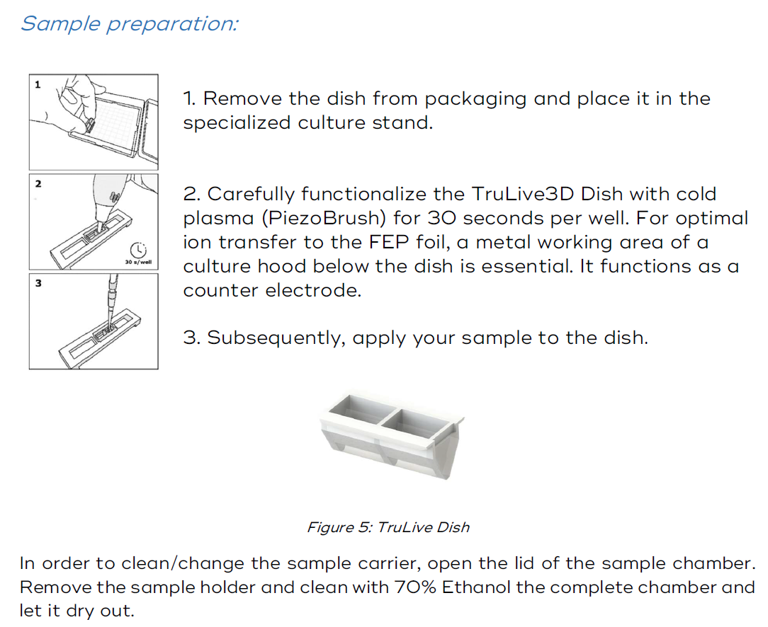
3. How to prepare a sample in the Bruker TruLive3D dishes
Below are the recommendations given by Bruker about sample preparation:

 \
\
No Comments