Single Objective Light Sheet Microscope (OPM)
Oblique Plane Microscope (OPM) Acknowledging the Arnold & Mabel Beckman Foundation for their support in financing the equipment for this instrument.
- Specifications and primary uses of the OPM
- Using the OPM
- Sample preparation for the OPM
- Setting up an experiment on the OPM
- OPM Data processing in Fiji
Specifications and primary uses of the OPM
Single Objective Light Sheet Microscope
The Single Objective Light Sheet Microscope is also referred to as Oblique Plane Microscope or OPM. It is a custom design Light Sheet microscope that enables high resolution and can be used with standard coverslipped samples. There is virtually no sacrifice in numerical aperture (NA) or loss in resolution - unlike most light sheet microscopes.
The OPM therefore presents a strict improvement over standard confocal microscopy. It has the sectioning capability and reduced photo-toxicity of light sheet microscopes while preserving image quality.
The animation below illustrates how it works.
High_NA_single-objective_light-sheet_scan.mp4
For more technical information on the OPM design, check this open source paper.
The Beckman Center Single Objective Light Sheet Microscope
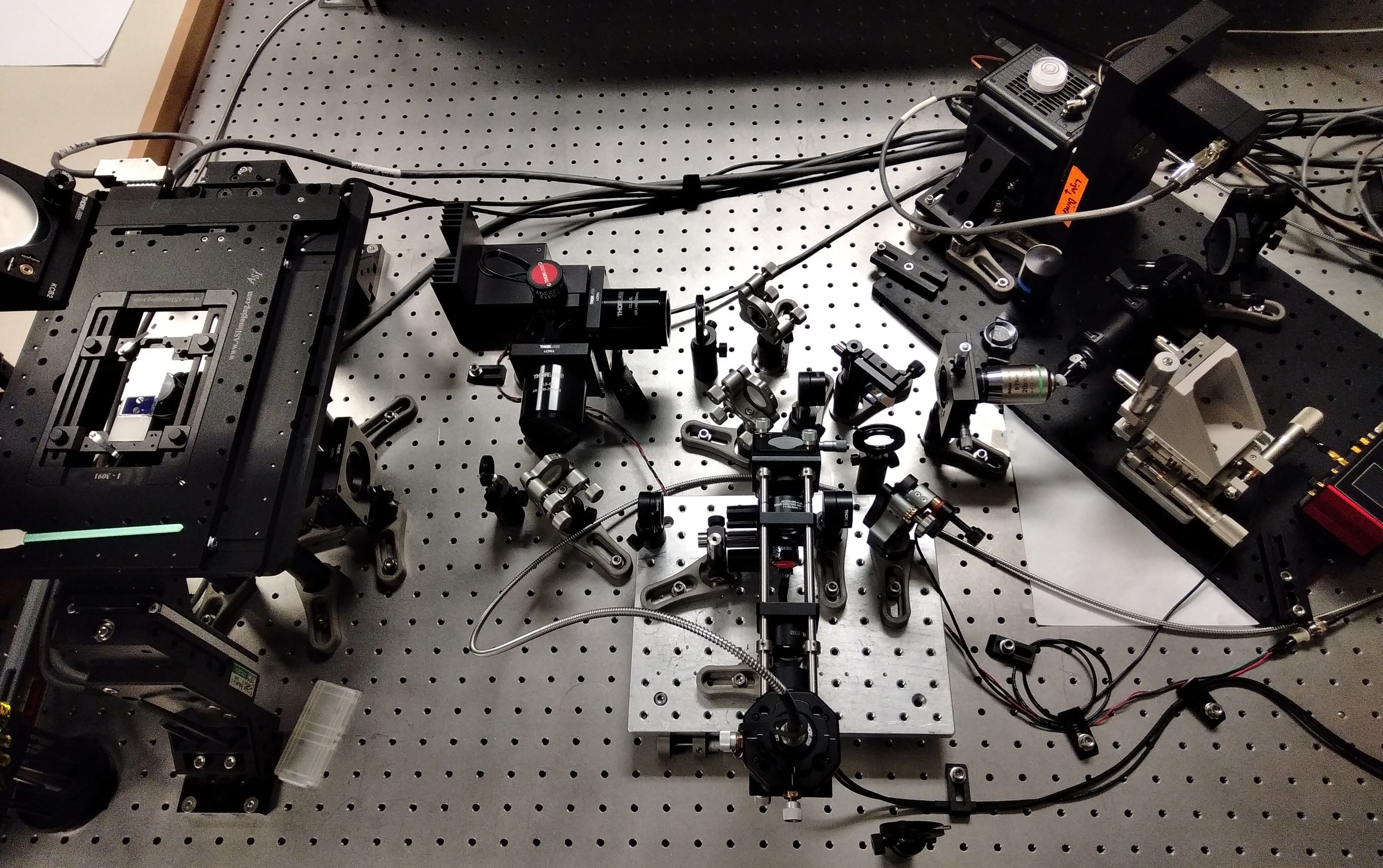
- Primary objective: Olympus 20X 1.0NA water dipping
- Secondary objective: Nikon 20X 0.75NA
- Tertiary objective: AMS-AGY V1.0 "Snouty"
- Hamamatsu Fusion sCMOS camera
- ASI FTP2000 XYZ stage
- 4 Channels: 405, 488, 561, 640
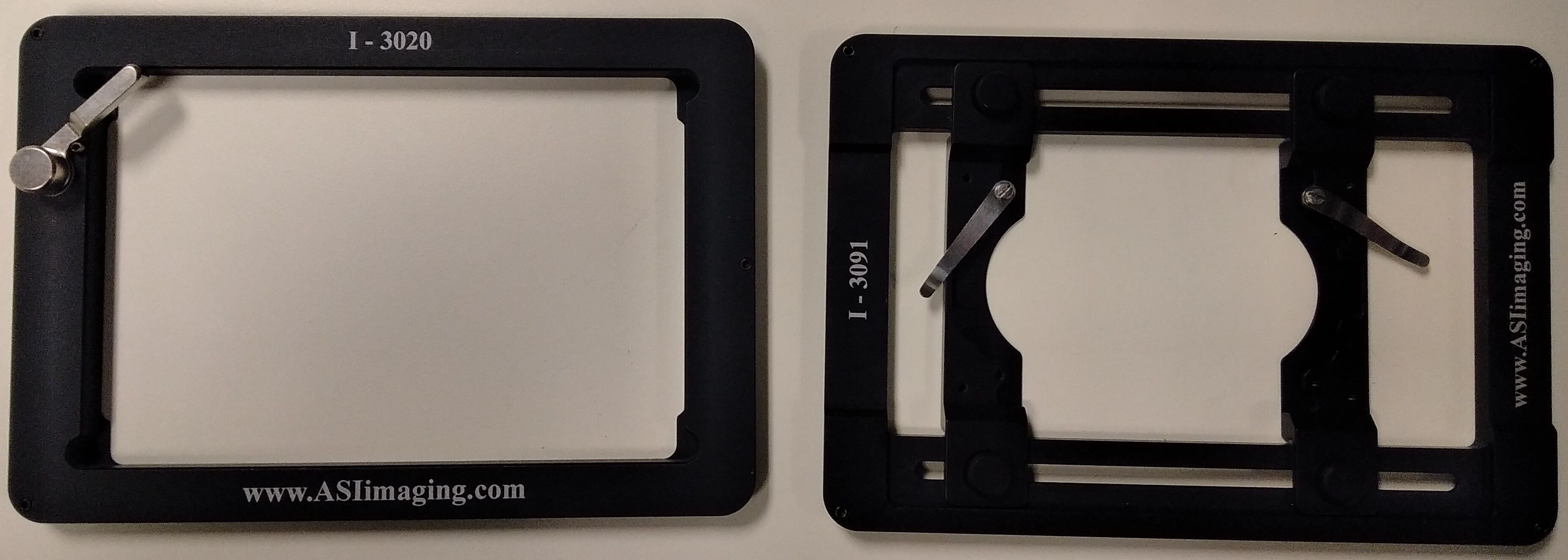
The sample stage has several sample holders for slides, round dishes and multi-well plates.
Primary uses
The size range of the samples is roughly from single cells to the bottom layer of small drosophila or zebra fish embryos and small organoids. Typically the OPM is used for 1 to 3 cells deep into the sample.
While this limits the variety of sample that can be imaged on the OPM, the flexibility of sample preparation is a considerable advantage. The fact that is uses the same dishes as other standard commercial microscope allows for straightforward comparison of images between a confocal microscope and the single objective light sheet microscope.
Examples
Some imaging examples using the OPM technology by The Dean Lab at UT South-Western, Dallas Texas:
- Biological imaging of clathrin-mediated endocytosis, vimentin and membrane dynamics
- Natural killer cell mediated cytotoxicity
- Imaging in biological microchannels - microtubules and nuclear shielding
- High-speed imaging of calcium transduction and cytoplasmic flows
- Simultaneous volumetric imaging and optogenetic stimulation
- Large field of view imaging of cortical neurons and ventral furrow formation
- Tissue-scale imaging
For further details and reference, the original article "A versatile oblique plane microscope for large-scale and high-resolution imaging of subcellular dynamics" can be found here.
Below is an example of what the OPM can do for tissue scale imaging:
"In addition to the rapid laser scan/descan illumination geometry, OPM is also compatible with a sample scanning acquisition format that is essentially field of view unlimited. Indeed, by combining scan optimized equipment with fully automated fluidic handling, it is possible to image ~1 cm2 of a thin tissue in less than 45 min per color and perform biochemistry, such as sequential multiplexed labeling. To demonstrate this, we imaged an entire 30-micron thick slice of coronal mouse brain tissue (Figure 9A, Video 15) labeled with the nuclear marker DAPI. Within these data, even small features like nucleoli are clearly resolved from both lateral and axial viewing perspectives throughout the entire ~6×8 mm tissue slice (Figure 9B and C). Likewise, we also imaged a ~ 4×14 mm slice of 12-micron thick human lung tissue labeled for nuclei, angiotension-converting enzyme 2 (ACE2) mRNA, and surfactant protein C (SFTPC) protein (Figure 9D). Here, characteristic histological features, including bronchiole, alveoli and vasculature, are readily visible, albeit with molecular contrast and sub-cellular resolution (Figure 9E,F and G, and Video 16). Quantification of molecular expression within this tissue section provides spatial information on ~20,000 cells, and verifies our previous limited quantification of ACE2 expression in alveolar epithelial type II cells using confocal microscopy (Muus and Luecken, 2020). Indeed, because we were not sterically restricted by the orthogonal illumination and detection geometry (Figure 9—figure supplement 1), the lateral dimensions of this human lung specimen were 8- and 1.5-fold larger than those of the biggest sample imaged with lattice light-sheet microscopy (Gao et al., 2019). However, in the third dimension, lattice light-sheet microscopy has in principle a 6.7x larger reach (2 mm working distance of the typically employed NA 1.1/25X detection objective compared to 300 microns working distance of our primary objective). In practice, optical aberrations limit high-resolution light-sheet microscopy to depths of a few hundreds of microns, even for highly transparent samples. Furthermore, our approach is fully compatible with automated fluid exchange, which is increasingly important for projects like the Human Cell Atlas that necessitate iterative imaging approaches for spatial -omics of RNAs and proteins at the single-cell level throughout entire tissues (Chen et al., 2015)."
Using the OPM
The Single Objective Light Sheet Microscope is a custom-designed microscope and is a VERY sensitive instrument. It is very easy to bump components out of alignment.
For this reason, you will not be allowed to use it in autonomy. There are no trainings provided at the moment, all your experiments will require the presence and supervision of the Beckman Center staff.
Your path to imaging your samples with the OPM
- Email the ticketing system at biof-imaging@colorado.edu and request a consultation with the Light Sheet Specialist.
- Book an appointment using the Microsoft booking link you will receive in the email from the ticketing system.
- Come prepared to the consultation with a clear idea of what your imaging needs are.
- You will then schedule a time on the instrument with the Beckman Center staff for a general overview of the instrument, the software and the Data generated. You will do some preliminary imaging then if you have your sample ready. This session will be similar to a classical training except you will only watch.
- Immediately after, or at another time, you will learn to process and visualize the data you acquired.
- Later on, always inquire with the Beckman Center staff about availability for scheduling your next imaging session.
Sample preparation for the OPM
Good news: Traditional sample preparation!
The OPM sample holders are similar to that of other commercial microscopes. Slides, round dishes and multi-well plates can be used. There is nothing different for your sample preparation so far.

Setting up an experiment on the OPM
This page is a guide to set-up an experiment with the OPM.
All experiments on the OPM will be done under the supervision of the Beckman staff and you will get help with processing the data to make it ready to analyze.
Turning the system on
Inserting the sample on the microscope
Turning Navigate on
Finding the sample and an area of interest
Setting up a basic experiment
How is the Data saved
OPM Data processing in Fiji
OPM Data
The data generated by the OPM is in TIFF format. Whether you acquire a single image or a z-stack, you will get a single TIFF file containing all the images.
The software also saves an "experiment" file in Yaml format that contains all the experiment settings as well as a "waveform_constants" file in Yaml format that contains the waveform settings. These files can be opened by different applications such as notepad, etc. If you have Visual Studio on your computer, it is a great application to open the file in a structured way. See example below.

Data processing with Fiji/ImageJ
Of course, you are welcome to use any software/method you like to process, analyze and render your data. The TIFF file format should make it easy to open by most softwares.
However, until further development of the instrument, the acquired data is skewed due to the angle of the beam scanning to capture the z-stack. Before visualizing it, you will need to perform a shearing operation to "de-skew" the data.
THE BECKMAN CENTER STAFF WILL DO THIS FOR YOU!!
But if you are curious about how to do it yourself, here are the steps:
There is a way to do this using Fiji/ImageJ. It requires a GPU and CLIJ installed on ImageJ.
- How to install CLIJ on ImageJ:
- Follow the steps indicated on this github page. A Wiki page about CLIJ will be available soon as well.
- Shearing the data:
- Drag and drop the Fiji macros code in Fiji (provided to you by Beckman Center staff).
- Go in your experiment folder and copy the TIFF file of your z-stack into an empty separate folder (call it "Raw Data" to avoid confusion)
- On the code, update the angle values if different than 40 degrees, and the z step size. The xy pixel size should not change for this microscope.
- Click Run and when prompted, select the TIFF file of your z-stack in the Raw Data folder.
- Rotate and visualize the data:
- Two more scripts are used to visualize and rotate the data to top view and side view. They can be provided to you by the Beckman center staff.
In practice, once your experiment is finished, the Beckman Center staff will give you the processed data. However, if you are an enthusiastic Fiji user and want to further process and analyze you data in Fiji you might find it useful to be able to perform the de-skewing operations yourself to tweak them to your needs.